NEW VISION FUNDUS – New Vision FUNDUS

NEW VISION FUNDUS

NewVision Fundus U.S. FDA approved ophthalmic medical image processing system

New Vision Fundus is the best choice for professional Medical Imaging.
Europe’s #1 top selling, U.S. FDA approved ophthalmic medical image processing and digital storage software!
New VISION Fundus

Europe’s #1 top selling, U.S. FDA approved ophthalmic medical image processing and digital storage software!


FDA

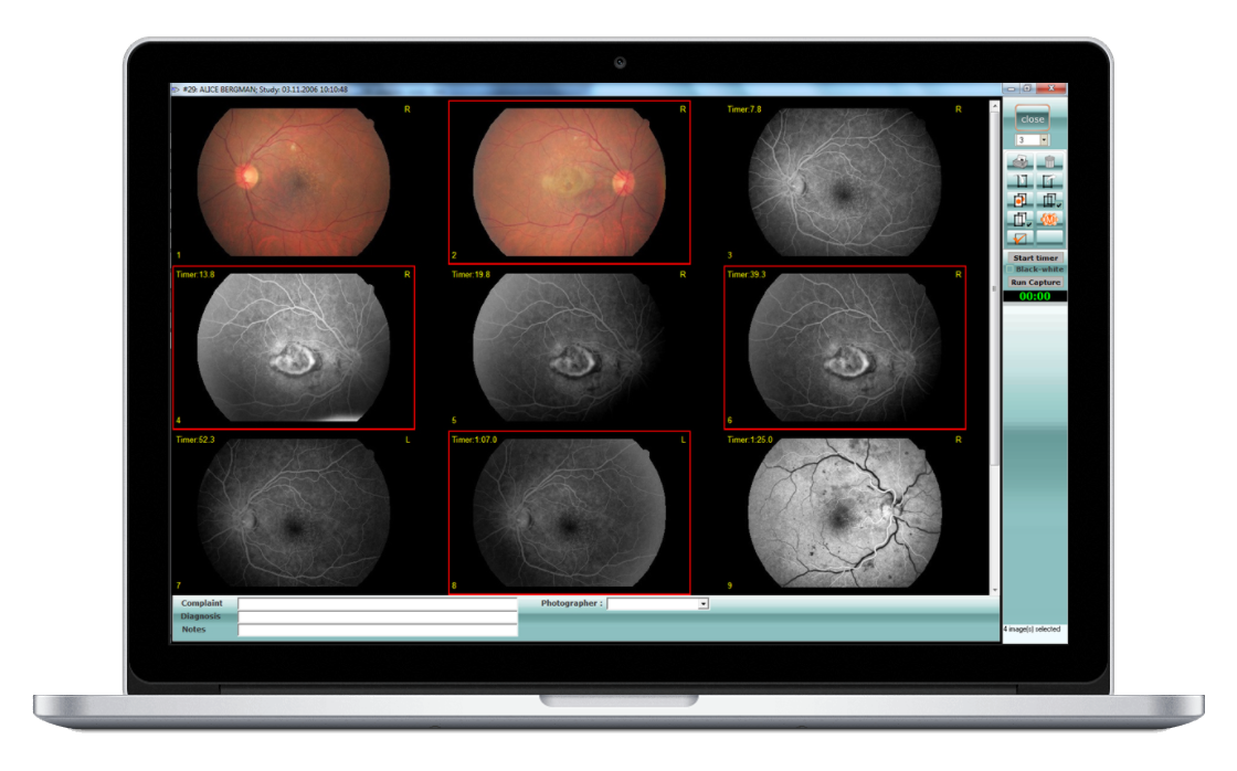
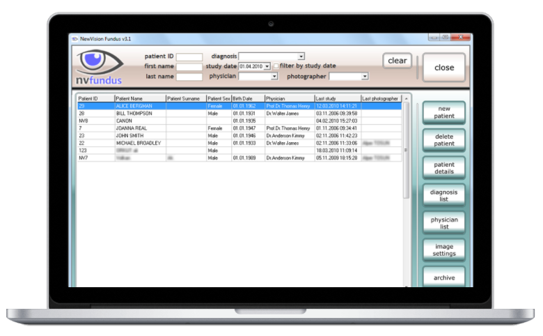
Fundus Fluorescein Angiography (FFA) and ICG angiography are made simple by the easy-to-use NewVision software. Patient EMR, archive, printing quality reports, and burning images to DVDs has never been so simple.

CE

We declare with our EC Declaration of Conformity that our New Vision Fundus image processing functions, measurement, and documentation Software meets the essential health and safety requirements of the European Community (EC) directives and standards issued by the European Community (EC).

Functions

Various image processing functions provide tools for diverse imaging tasks such as visualization, enhancement, measurement, and documentation.

Modular architecture

Modular architecture of New Vision allows presentation of a custom system that will fit almost any Digital Imaging equipment and will satisfy any customer’s needs.

Cameras and software

NewVision integrates automated microscopes, digital cameras and software and provides convenient means for Digital Image acquisition, storage, manipulation, and analysis, as well as data exchange and data backup.

NEW VISION FUNDUS

Enhancement Technology in Fundus Images The retinal fundus image plays an important role in the diagnosis of retinal diseases. Detailed information of the retinal fundus image, such as small vessels, microaneurysms, and exudates, may be of low contrast, and retinal image enhancement often helps to analyze retinal fundus image-related diseases. Image enhancement can be applied to any fundus image taken thanks to the “Enhancement” function (image enhancement function) in New Vision Fundus. A new image can be saved by applying the desired image enhancement process to the images transferred to the New Vision Fundus. The “Enhancement” functions help the doctor to examine the fundus images taken with the ophthalmic camera in more detail. According to the feedback we received from our customers, it seems that the “Enhancement” functions benefit both doctors and patients. Thanks to the image enhancement process, images that are too dark to be evaluated are improved and brought to a level that can be analyzed by the doctor.


Before
After

Haze Enhancement Button

Before
After

Cataract FA Enhancement Button

Before
After

Color Enhancement Button

Before
After

Automatic Enhancement Button

Before
After

Automatic Enhancement (Medium) Button

Before
After

Automatic Enhancement (Aggressive)

URAL Telekom

Our Company

  • NewVision Fundus Imaging Suite was created in 2003 for Canon Ophthalmic microscopes. As the technology and opthalmic equipments developed our image processing software reaches its recent level “New Vision Fundus”.
  • URAL Telekom also develops Ultrasound PACS, Analysis and Storage software for Obstetrics, Perinatology, Cardiology and general Ultrasound imaging procedures.
  • URAL Telekom also specializes in Software Performance Optimization, Legacy Software Migration, Test Automation Consulting services.
  • The company gains its strength from the innovative approach and cooperative culture continued since its foundation, besides its economical solution strategy and high level of client satisfaction.

URAL Telekom


New VISION Fundus

U.S. FDA APPROVED OPHTHALMIC MEDICAL IMAGE PROCESSING AND DIGITAL STORAGE SOFTWARE

Fundus Image Analysis and Enhancement

  • Automatic image enhancement;
  • Image editing and manipulation;
  • Mosaic Tool;
  • Transparent image Overlay and comparison;
  • Image annotation;
  • Image-to-image registering and concurrent drawing;
  • Cup-to-disk ratio measurement;
  • Automatic PDT drawing and analysis;
  • Magnifying Glass Tool, Automatic Zoom Tool, Negative Image Tool and many more;
  • Recovery of low-quality images;
  • Endothelial Cell analysis (optional);

DICOM Support

  • DICOM Storage Server
  • DICOM Modality Worklist
  • DICOM Modality Worklist Find
  • DICOM Image processing support (optional);
  • DICOM Confirmance Statement

Fundus Camera Image Capture

  • Automated image acquisition from any type of digital camera and fundus camera;
  • Easy image drag-and-drop inserting;
  • Fundus Fluorescein Angiography (FFA);
  • ICG angiography;
  • Color images;

Additional features

  • Web-based analysis support;
  • Efficient backup and restore mechanism;
  • Direct burning to CD and DVD;
  • Customizable printout support;
  • Easy jpg Export Tool for Power Point Presentations and Export to any destination you choose;
  • Network with multiple workstations for viewing in other locations;
  • Video Storage and Playback support (optional);
  • Support for MSWord, MSExcel and AdobePDF documents (optional)

EMR and PACS Systems integration

  • Integration with all standard systems
  • DICOM integration

New VISION Fundus DICOM Server

Overview New Vision DICOM Server is a complete DICOM Solution enabling seamless integration of all types of DICOM-enabled automated microscopes and digital cameras with New Vision Medical Imaging Software, leading ultimately to a complete Hospital PACS system. New Vision DICOM Server offers full-featured DICOM support and functionality.


NewVision Fundus DICOM Server Features

  • DICOM 3.0 Compatible;
  • Includes a DICOM Server SCP;
  • Uses fast MySQL Database;
  • Fully supports Lossless Jpeg Images;
  • Creates DICOM objects from any image;
  • Archive images to DICOM servers;
  • Query other DICOM servers and retrieve images;
  • Web access (optional);

DICOM Conformance Statement

NewVision Fundus DICOM Server is a Level 2 (Full) Service Class Provider:

  • Storage Service Class – Level 2 (Full);
  • Related General SOP Class – Level 2 (Full);
  • Signature Support – Level 3;
  • Lossless Jpeg support – 1.2.840.10008.1.2.4.70, 1.2.840.10008.1.2.4.57;
  • After a successful C-STORE command a SOP instance is stored in a local DICOM Directory
  • SOP instance is saved to a database together with patient and study information.
  • SOP Instances are accessed via C-GET protocol or using New Vision Imaging Software;
  • SOP Instances are stored indefinitely, until deleted by user or moved to a permanent storage (Backup);
  • Full implementation of DIMSE-C protocol.
  • Detailed DICOM Confirmance Statement